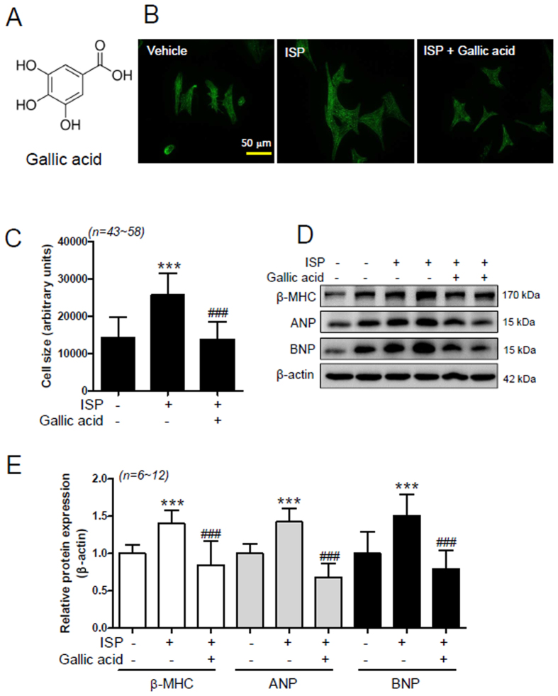
Figure 1. Gallic acid pretreatment suppresses cardiomyocyte hypertrophy in vitro.
(A) Chemical structure of gallic acid. (B–E) Rat neonatal cardiomyocytes were pretreated with vehicle or 100 μmol/L gallic acid for 30 min and then coincubated with 10 μmol/L isoproterenol (ISP) for 12 h or 24 h. (B) Stress fiber was evaluated by immunocytochemistry with anti-sarcomeric α-actinin antibody (scale bar = 50 μm). (C) Cell size was calculated by measuring the cell surface area (n = 43~58 per group). Data are presented as the means ± SD of 3 independent experiments. ***P < 0.001 versus vehicle; ###P < 0.001 versus ISP + vehicle. (D) Cell lysates were subjected to SDS-PAGE and incubated with anti-β myosin heavy chain (β-MHC), atrial natriuretic peptide (ANP), and brain natriuretic peptide (BNP). β-Actin was used as a loading control. (E) β-MHC, ANP, and BNP protein amounts were quantified by densitometry. Data are presented as the means ± SD of 3 independent experiments. ***P < 0.001 versus vehicle; ###P < 0.001 versus ISP + vehicle.

