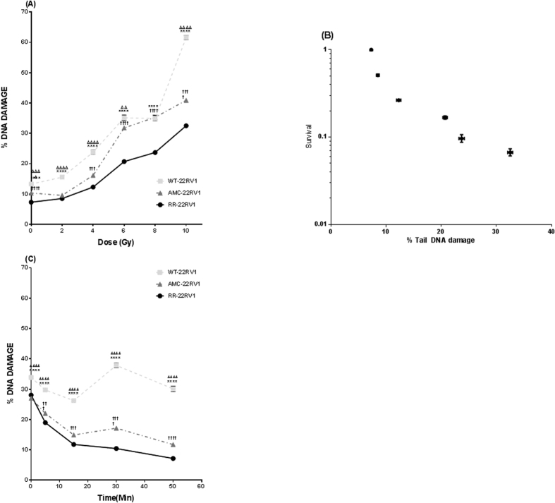
Figure 3. Increased proliferative potential following radiation exposure is associated with reduced levels of DNA damage.
(A) DNA damage in response to treatment with a range of radiation doses (0–10 Gy) in each of the three cell lines as measured by alkaline comet assay. N = 3 independent experiments. (B) Correlation between clonogenic survival and % tail DNA damage in 22Rv1-RR cells. (C) DNA repair capabilities of the 22Rv1-RR, WT and AMC cell lines. Initial damage and repair up to 50 min post irradiation with 8 Gy is measured using the alkaline comet assay. n = 5 independent experiments. DNA damage was calculated using % tail DNA for each comet, 400 comets were scored on two slides/4 gels per replicate and SEM calculated. *refers to significant differences in the RR cell line vs WT control. †refers to significant differences between RR and AMC cells. Δ refers to significant differences between AMC and WT control cells. *P ≤ 0.05, **P ≤ 0.01.

