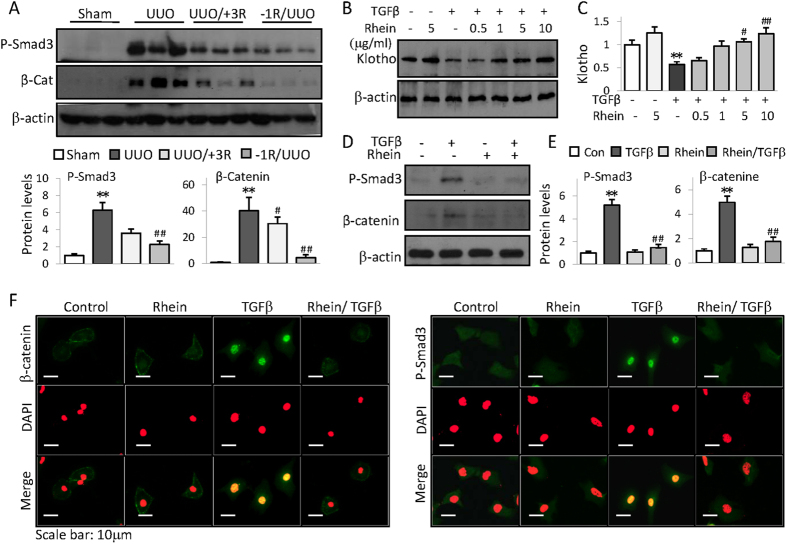
Figure 2. Rhein reverses Klotho suppression and inhibits profibrotic cellular signaling.
(A) Renal expressions of phosphorylated Smad3 and β-catenin from Sham, UUO, or UUO mice treated with Rhein 3 days after (UUO/+3R) or 1 day before UUO (−1R/UUO) were assayed by Western blot (3 samples from each group). Quantifications were underneath the figures. (B) HK2 cells were treated with TGFβ in presence or absence of increasing doses of Rhein for 48 h. Klotho protein was assayed by Western blot. (C) Quantification of (B). (D) HK2 cell expressions of phosphorylated Smad3 and β-catenin when treated with Rhein (10 μg/ml) in presence or absence of TGFβ (5 ng/ml) for 4 h or 24 h, respectively. (E) Quantification of (D). Data were represented as mean ± SD of three independent experiments. *p < 0.05, **p < 0.01 versus Control or Sham, #p < 0.05, ##p < 0.01versus UUO or TGFβ treatment. (F) Representative immunofluorescent staining of phosphorylated Smad3 and β-catenin from HK2 cells treated with Rhein in presence or absence of TGFβ for 4 hours. The cells were also stained with DAPI (middle panel) and merged with P-Smad3/β-catenin images (lower panel). The original blue nuclear DAPI staining was converted to red so that the nuclear green P-Smad3 or β-catenin staining was shown in yellow color on merged images for easy recognition.

