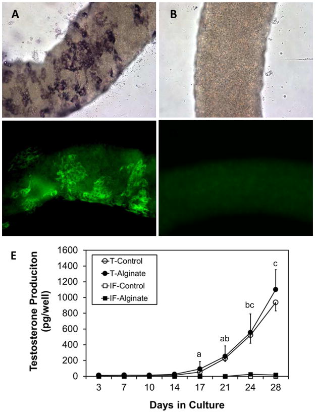
Figure 2.
Effect of encapsulation on the development of functional Leydig cells in vitro. Alginate encapsulated seminiferous tubules were stained for 3β-HSD (A, B) or CYP11A1 (C, D). Clusters of 3β-HSD (A) and CYP11A1 (C) positive cells were seen after 4 weeks in culture, but not before culture (B, D). (E) Testosterone production by cultured seminiferous tubules. Whether encapsulated or not, the cultured tubules (T) produced testosterone by 4 weeks, whereas intersitium (IF), whether or not encapsulated, produced no testosterone. Data are expressed as mean ± SEM of three separately conducted experiments. Groups with shared letters were not significantly different at P≤0.05. Groups without letters had undetectable testosterone levels.

