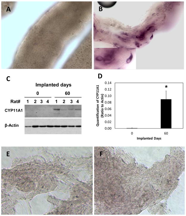
Figure 4.
Formation of steroidogenic cells in implanted seminiferous tubules in vivo. (A) Seminiferous tubules before their implantation, showing absence of 3βHSD-positive cells. (B) 3βHSD-positive cells were found in the seminiferous tubules two months after implantation. (C) Western blot of CYP11A1 before (0 days) and 60 days after tubule transplantation into four castrated rats. Actin served as a loading control. (D) Quantification of CYP11A1 at 0 and 60 days after tubule transplantation. *Significantly different compared to day 0 at P≤0.05 (N=4). (E, F) No 3βHSD-positive cells were seen associated with interstitium before (E) or two months after (F) implantation.

