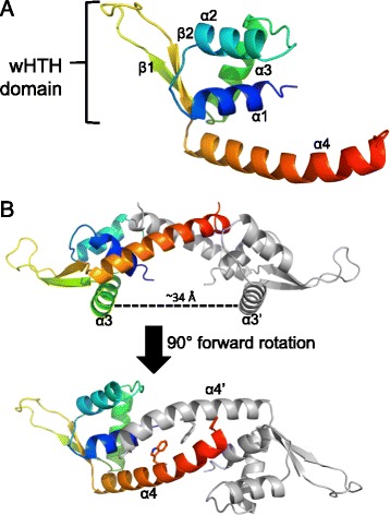
Fig. 2.

Overall structure of cdPadR1. a Ribbon representation of cdPadR1 monomer with a rainbow color gradient from the N-terminus (blue) to the C-terminus (red). Alpha helices and β-sheets are labeled numerically. The winged helix-turn-helix (wHTH) DNA binding domain is indicated. b The cdPadR1 structure is shown perpendicular to the two-fold axis of symmetry with the DNA recognition helices indicated (α3/α3′). The dimer mate is shown in gray. Distance between α3/α3′ was estimated in PyMOL [22]. Another view is shown after a 90° forward rotation which results in a view along the two-fold axis facing α4/α4′ with the conserved W residue (sticks)
