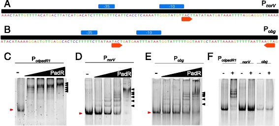
Fig. 6.

EMSAs of cdPadR1 binding the cdpadR1 promoter (PcdpadR1), a norV promoter (PnorV), and b obg promoter (Pobg). The predicted -10 and -35 sites are indicated in blue boxes above the sequence. Orange arrows indicate the inverted repeats TACT/AGTA. c-e Final PcdpadR1, PnorV, and Pobg (100 bp) concentration in the reaction was 0.1 μM. cdPadR1 was 5, 10, 20, and 40-fold excess over dsDNA. The minus (-) lane contains dsDNA and no cdPadR1. Shifted DNA-protein complexes are annotated with a black arrow and unbound dsDNA migration is marked with a red arrow. f For this EMSA, the final heparin concentration was 4-fold of the standard EMSA concentration used throughout this research. The + lane contains 40-fold excess cdPadR1 over dsDNA
