Fig. 2.

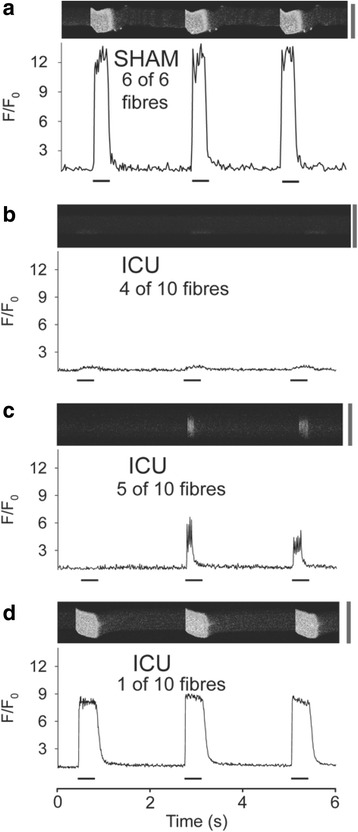
Ca2+ release from the sarcoplasmic reticulum is reduced and non-uniform in muscle fibers from ICU rats. Panels a-d show representative line scans of fluo-3 fluorescence from flexor digitorum brevis (FDB) fibers during three 70-Hz tetani at 2-s intervals. a Typical homogeneous free myoplasmic [Ca2+] ([Ca2+]i) transient during each of the three tetani seen in sham-operated (SHAM) rat fibers. b-d The [Ca2+]i transients observed in ICU rat fibers. b Ca2+ release is seen only at one edge of the fiber. c The [Ca2+]i transients do not occur in response to each tetanic stimulation and do not last throughout the whole period of electrical stimulation. d The [Ca2+]i transients occurred during each of the three tetani but were reduced compared to those in SHAM rat fibers. Fluorescence intensity (F) is expressed relative to the value at rest (F 0). Periods of electrical stimulation are indicated by the black bars underneath each trace. Calibration bar to the right of each line scan is 50 μm
