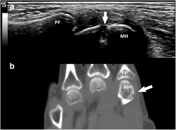
Fig. 1.

Bone erosions in a patient with rhupus syndrome. Longitudinal ultrasound image of the second metacarpophalangeal joint lateral area showing a grade-2 scoring ultrasound structural erosion (arrow) with sharp margins (a) corresponding to a grade-2 rheumatoid arthritis magnetic resonance imaging score erosion (arrow) visible in the computed tomography coronal scan of the hand (b). MH second metacarpal head, PP second proximal phalanx
