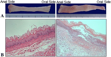
Fig. 1.

Gross specimen (a) and hematoxylin and eosin (H&E) staining of esophageal mucosa (b) 72 h after surgery in sham operation group (left) and RE model groups (right); normal esophageal epithelial and a small quantity of inflammatory cells were seen in sham operation group; Lower ulcer with peripheral congestion and swelling was seen in RE model groups 72 h after surgery. H&E staining showed that epithelial defects and inflammatory cells infiltration. Several neutrophils, eosinophils, and monocytes were seen under high magnification
