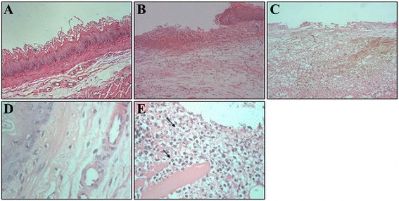
Fig. 3.

H&E staining of esophagitis under microscopy. a Normal esophageal epithelial with stratified squamous epithelium (H&E staining, 100×); b Inflammatory cells were seen under high magnification (H&E staining, 100×); c Epithelial defects in acute RE models. d Numerous neutrophilic granulocytes, eosinophilic granulocytes, and monocytes were seen under high magnification (H&E staining, 400×). e Some mucosal disruptions occurred including ulcer, necrosis, and bleeding (H&E staining, 400×). The arrow represents neutrophils
