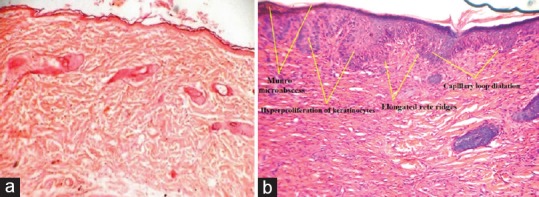
Figure 1.

Longitudinal histological sections of mouse skin (H and E, ×40), (a) section of normal mouse skin and (b) section of complete Freund's adjuvant- and formaldehyde-treated mouse skin. (The arrows are pointing at Munro's microabscess, hyperproliferation of keratinocytes, elongated rete ridges, and capillary loop dilatation)
