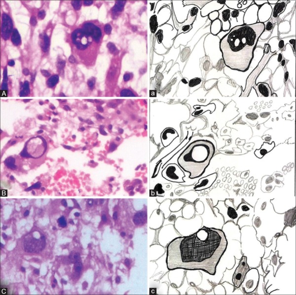
Figure 1.
(A) Photomicrograph of a neoplastic cell showing lochkern nucleus (H&E stain, ×400), (a) hand-drawn illustration showing Lochkern nucleus. (B) Photomicrograph of a neoplastic cell showing Ringkern nucleus (H&E stain, ×400), (b) hand-drawn illustration showing Ringkern nucleus. (C) Photomicrograph of a neoplastic cell showing Kerbkern nucleus (H&E stain, ×400), (c) hand-drawn illustration showing Kerbkern nucleus

