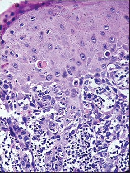
Figure 3.

Photomicrograph shows moderate dysplasia in the case number 6 at the time of diagnosis of oral lichen planus (H&E stain, ×400)

Photomicrograph shows moderate dysplasia in the case number 6 at the time of diagnosis of oral lichen planus (H&E stain, ×400)