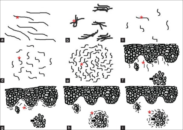
Figure 2.
Patterns of elastic fibers. The figure depicts line diagrams of various patterns exhibited by elastic fibers in varying grades of oral squamous cell carcinoma. (a) Long, thin and straight elastic fibers present singly, (b) short, thick and wavy elastic fibers present in bunches; density of elastic fibers, (c) scanty, (d) moderate, (e) abundant; orientation of elastic fibers in relation to overlying epithelium (f) perpendicular, (g) parallel; orientation of elastic fibers in relation to tumor islands (h)= parallel, (i) haphazard. Asterick = Elastic fiber, Arrow = Tumor island

