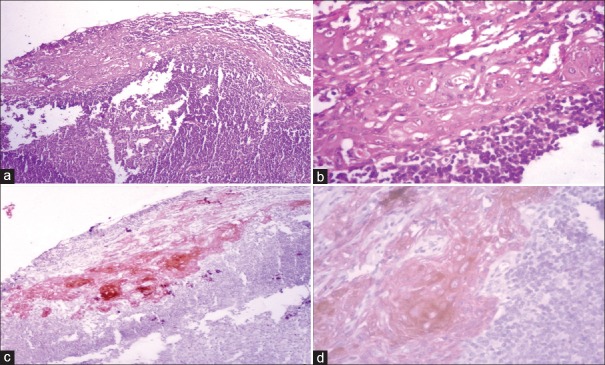
Figure 2.
(a) Lymph node of moderately differentiated squamous cell carcinoma revealed reactive hyperplasia at low-power (H&E stain, ×100); (b) high-power view of the same area revealed epithelial-like cells (H&E stain, ×400); (c) immunostaining with pan-cytokeratin (AE1/AE3) depicted metastatic deposits at low power (IHC stain, ×100) which were confirmed to be micrometastasis at high power (IHC stain, ×400)

