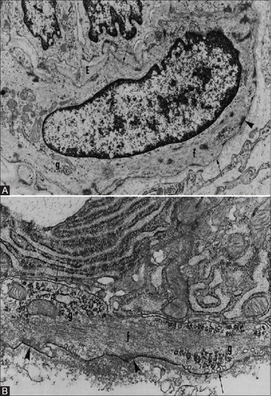
Figure 5.

Comparison between myoepithelial cells and smooth muscle cells. (A) Smooth Muscle cell (B) Myoepithelial cell. There are groups of filaments (f); dense bodies; attachment plaques (arrowheads); caveolae (arrows) in the plasmalemma, that are more numerous basally; basal laminae; and focal collections of glycogen particles (g) (Redman R.S. Myoepithelium of Salivary Glands. Microscopy research and technique 2725-45 [1994])
