Figure 7.

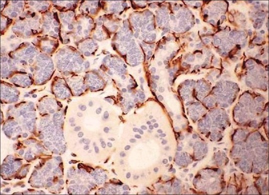
Demonst ration of myoepithelial cells using immunohistochemistry marker cytokeratin AE1/AE3 (IHC stain, ×400) (Courtesy: http://www.pathologyoutlines.com/topic/salivaryglandssuperpage.html [last viewed on 10/04/2015])

Demonst ration of myoepithelial cells using immunohistochemistry marker cytokeratin AE1/AE3 (IHC stain, ×400) (Courtesy: http://www.pathologyoutlines.com/topic/salivaryglandssuperpage.html [last viewed on 10/04/2015])