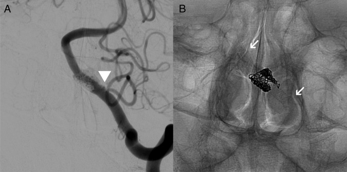
Figure 3.
Catheter cerebral angiogram, left vertebral artery injection, posteroanterior projections. (A) Subtracted angiogram with minimal filling of the sac proximally after coil assisted flow diversion (arrowhead). (B) The stent construct, with arrows marking the proximal and distal ends of the stent.

