
Fig. 1.
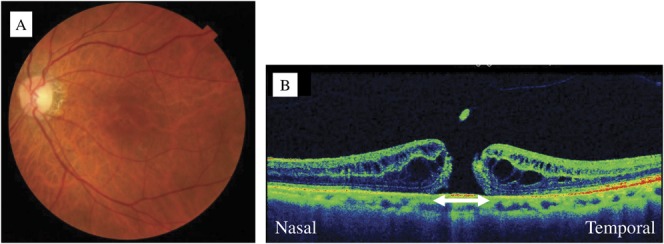
A. A preoperative fundus photograph of the left eye. B. An optical coherence tomography image shows a stage III MH. The maximal diameter of the MH is 351 μm (arrow).
Official websites use .gov
A
.gov website belongs to an official
government organization in the United States.
Secure .gov websites use HTTPS
A lock (
) or https:// means you've safely
connected to the .gov website. Share sensitive
information only on official, secure websites.
A. A preoperative fundus photograph of the left eye. B. An optical coherence tomography image shows a stage III MH. The maximal diameter of the MH is 351 μm (arrow).