
Fig. 3.
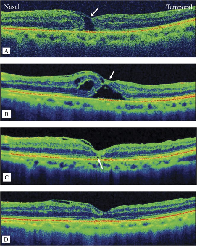
Postoperative optical coherence tomography images. A. at postoperative Week 1, the macular hole (MH) remains open under the covered internal limiting membrane (ILM) flap (arrow). B. at postoperative Week 3, the MH edges form a bridge beneath the ILM flap. C. at postoperative Week 4, the MH has closed leaving a partial defect in the inner segment/outer segment line (arrow). D. at postoperative Week 5, the full thickness of the fovea has recovered.