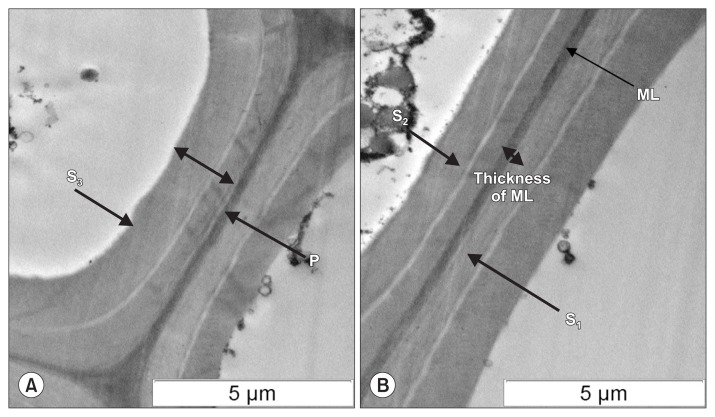
Fig. 3.
Transmission electron micrograph of ultrathin sections of oil palm seedlings cell wall after being stained with uranyl acetate and lead citrate at a magnification (× 6,000). (A) T9, healthy control. (B) T7, Ca/Cu/SA tissues. Ca, calcium; Cu, copper; SA, salicylic acid; S1, S2, and S3, secondary wall sub layers; P, primary wall; ML, middle lamellae.

