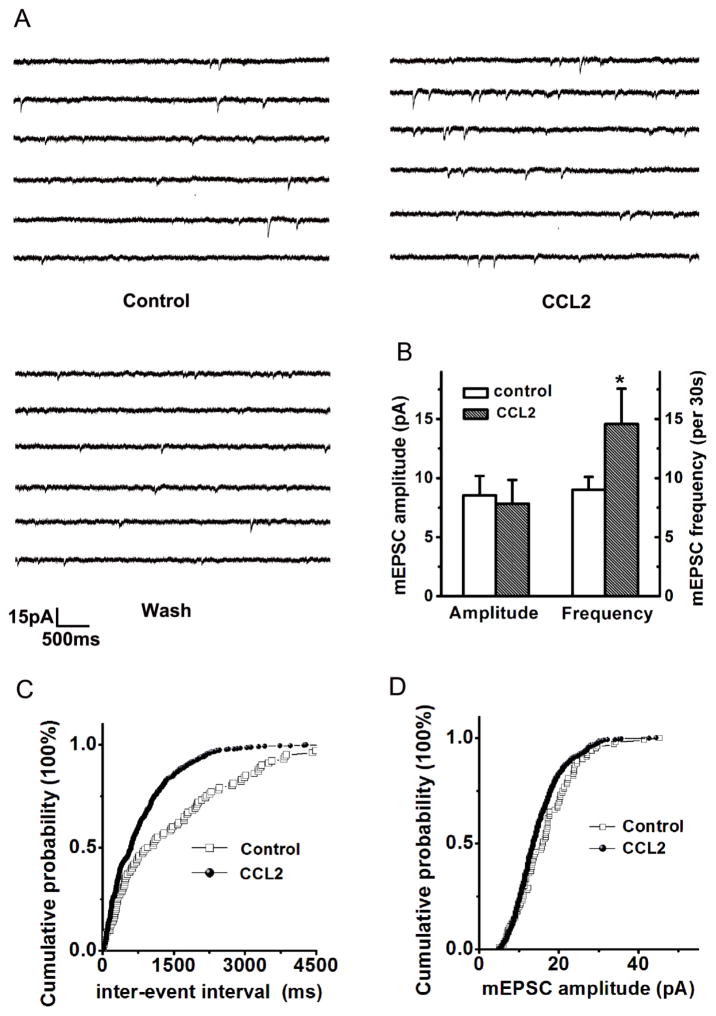
Figure 3. CCL2 Enhancement of EPSCs via a presynaptic mechanism.
Panel A shows the example traces of spontaneous mEPSCs recorded from a neuronal cell in the CA1 region of a hippocampal slice. Bath application of CCL2 significantly increased frequency of spontaneous mEPSCs without apparent effect on the amplitude (Panel B, n=9). Panel C exhibits cumulative distribution of mEPSCs inter-event interval (IEI) showing that CCL2 significantly decreased the IEI, indicating a significant increase in mEPSC frequency during bath application of CCL2 (p<0.05 vs control, n=9). A representative amplitude cumulative histogram is shown in panel D, showing no significant change (Kolmogorov-Smirnov-test) on mEPSC amplitude during bath application of CCL2.

