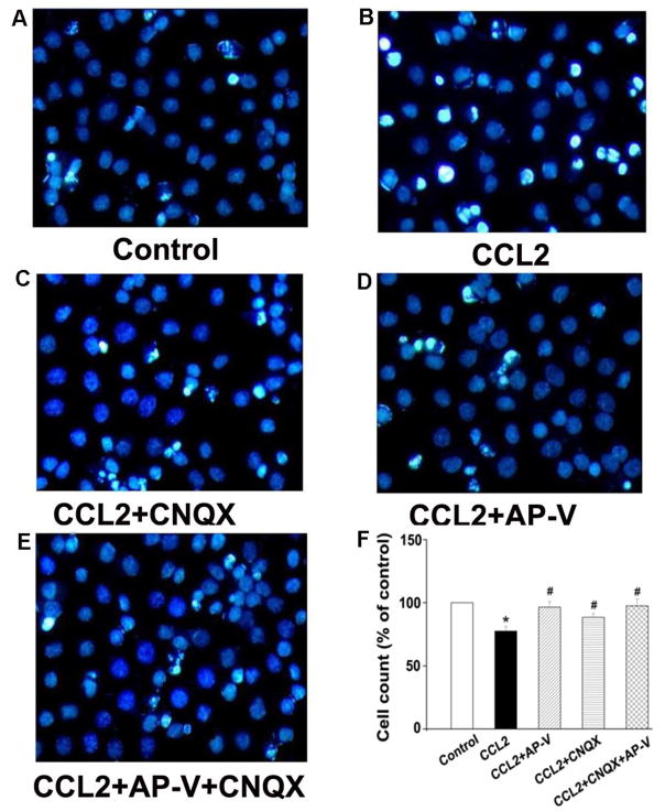
Figure 4. Attenuation of CCL2-induced neuronal injury by Hoechst staining.
Panels A–E are primary hippocampal neurons untreated (control) or incubated with CCL2, CCL2+CNQX, CCL2+AP-V, or CCL2+AP-V+CNQX as indicated. The injured cells were quantified after staining with Hoechst 33342. Survival rates were calculated by counting Hoechst-stained cells (bright blue) and total cells from five different visual fields in each dish contained cultured neurons are shown in panel F. CCL2 significantly decreased neuronal survival rates and the CCL2-induced reduction of survival rates were blocked by a NMDA receptor antagonist AP-V and by an AMPA receptor blocker CNQX as well. Addition of CNQX did not further improve the neuronal survival rates under existence of CCL2 and AP-V in the culture media, suggesting the CCL2-induced neuronal injury was largely mediated via NMDA receptors. * p<0.05 vs control, #p<0.05 vs CCL2. Experiments were done in three triplicates. Objective magnification: 40×

