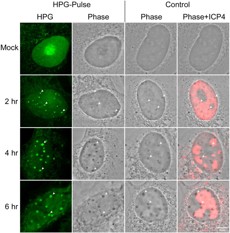
Fig 5. NPDs colocalise with phase-dense nuclear bodies induced by HSV.
Uninfected or HSV infected Vero cells were either untreated (i.e. standard media; control) or HPG pulse-labeled for 30 min at the times indicated, fixed and analysed by fluorescence (for newly synthesised proteins, green) and ICP4 (red) and by phase microscopy. Diagonal arrowheads indicate the colocalisation of NPDs with HSV-induced phase-dense nuclear domains. Identical phase dense bodies were formed in infected cells, localising to the periphery of replication compartments whether or not the cells were pulse-labeled with HPG.

