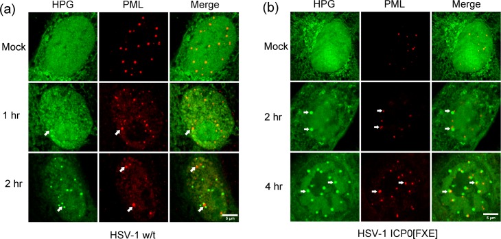
Fig 7. NPDs associate with PML domains in a spatially defined manner.
Vero cells were mock-infected or infected with w/t HSV-1 (A) or ICP0 RING-finger mutated HSV-1 strain FXE (B), then pulse-labeled with HPG for 30 min at 1 or 2 hr p.i., fixed and processed for newly synthesised proteins (green) and total PML localisation (red). Arrows point to the PML domains and are superimposed on the HPG channel to illustrate the juxtaposition of PML to a population of NPDs for the w/t and mutant viruses as discussed in the text.

