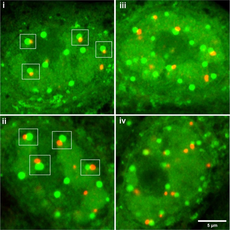
Fig 8. NPDs juxtapose to and overlap with PML domains in HSV-1 ICP0[FXE] infected cells.
Vero cells were infected with ICP0 RING-finger mutated HSV-1 strain FXE, pulse-labeled with HPG for 30 min at 4 hr p.i. and analysed as described in Fig 7. Several typical fields are illustrated. The clear spatial relationship between PML domains and NPDs are indicated in the white squares. While many persisting PML domains were associated with NPDs, NPDs were in excess of PML domains. Quantification data of NPD/PML association as discussed in the text are presented in S1D–S1E Fig.

