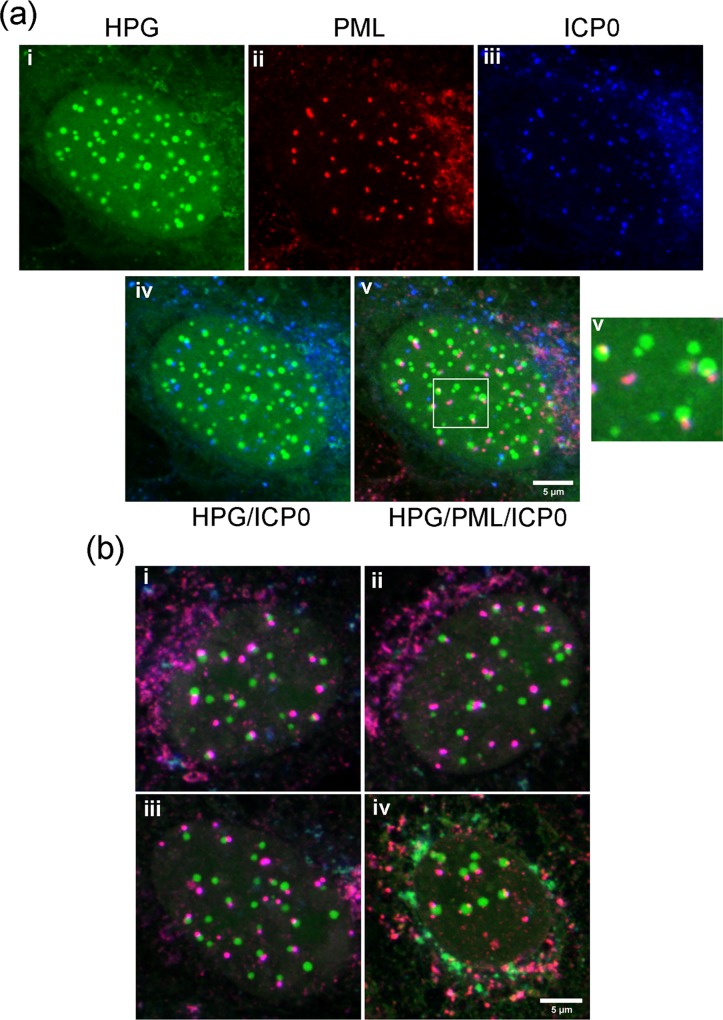
Fig 9. NPDs form adjacent to ICP0/PML domains.
(A) Vero cells were infected and processed as described in Fig 8, in this case for simultaneous localisation of newly synthesised proteins (i, green), PML (ii, red) and ICP0 (iii, blue). The higher magnification insert emphasises the juxtaposition of PML and ICP0 in relation to NPDs. (B) Typical examples of merged channels showing precise colocalisation of mutant ICP0 with PML (purple) and the juxtaposition with NPDs (green).

