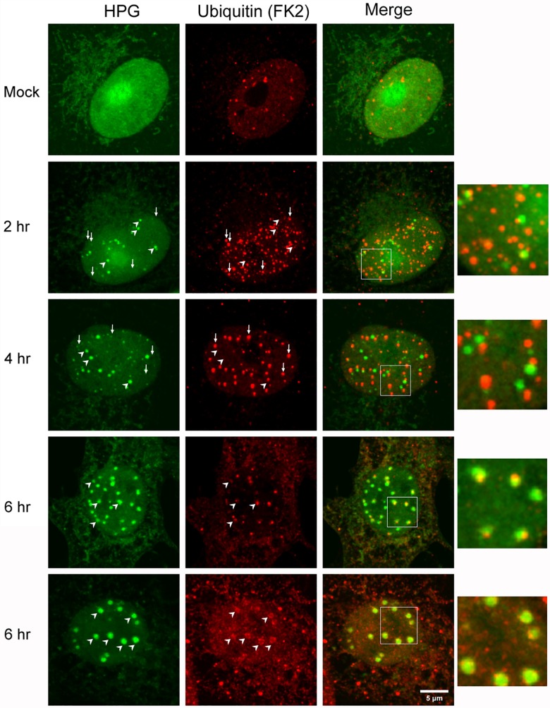
Fig 12. Polyubiquitinated species are recruited to NPDs at later stages of infection.
Vero cells were mock-infected or infected with HSV-1, pulse-labeled with HPG for 30 min at the times indicated, fixed and analysed for polyubiquitinated species (FK2 localisation, red) and newly synthesised protein (green). In mock infected cells HPG localised in a generally diffuse pattern with some nuclear la accumulation while polyubiquitinated species were found in a speckled diffuse nuclear pattern with variable numbers of discrete foci. NPD formation in infected cells is indicated by diagonal white arrowheads and early in infection NPDs show no colocalisation with FK2+ve foci. Conversely small vertical arrows indicate FK2+ve foci which show no obvious spatial relationship with NPDs. As described in the text (see also Fig 10) a subset of NPDs localised adjacent to, co-joining FK2+ foci (see inserts of the merged fields). Diagonal arrowheads later in infection show prominent co-localisation between NPDs and FK2+ve species, either as in example cell a, as co-joining asymmetric foci or frequently, as in example cell b, with virtually complete overlap FK2+ve species coating the exterior of the NPDs

