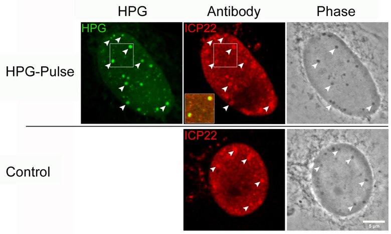
Fig 13. ICP22 localises to NPDs and phase-dense nuclear bodies.
Infected Vero cells were either untreated (standard media; control) or HPG pulse-labeled for 30 min at 2 hr p.i. (HPG), fixed and analysed by fluorescence (for newly synthesised proteins (green) and ICP22 (red) and by phase microscopy. Diagonal arrowheads indicate the colocalisation of NPDs with ICP22 as well as phase-dense nuclear domains. The inset shows the precise co-localisation of ICP22 with NPDs. The punctate localisation of ICP22, and its recruitment into phase dense bodies was independent of HPG pulse-labeling and also observed in the control infected cultures in the absence of HPG(Control).

