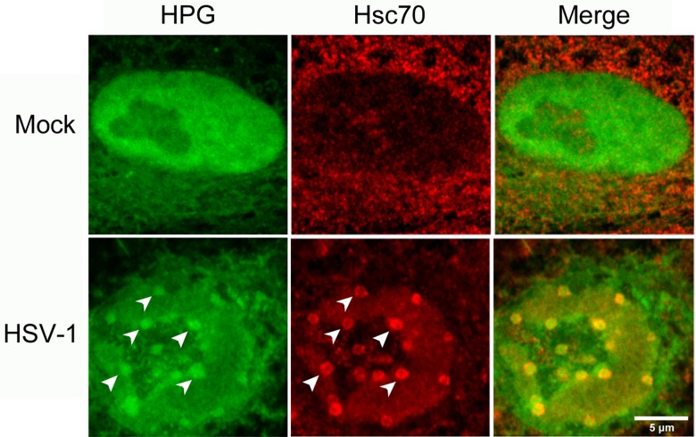
Fig 14. Fig Nuclear accumulation of cellular proteins synthesised prior to infection.
Uninfected Vero cells were pre-labeled with HPG (30 min pulse) followed by an infection with HSV-1 at an MOI of 10 for 4 hr (HSV-1) or mock infection (Mock). Cells were then fixed and stained for Hsc70, followed by click reactions. Cells containing Hsc70 foci were identified and the subnuclear localisation of newly synthesised proteins (green) and Hsc70 (red) are shown with diagonal arrowheads indicating the colocalisation of nuclear NPDs and Hsc70 foci as discussed in the text.

