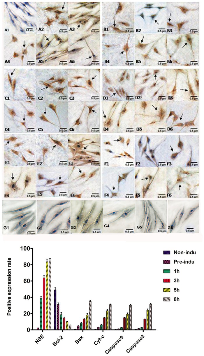
Fig 2. Expression of NSE, Bcl-2, Bax, Cyt-c, caspase-9, and caspase-3 during the process of ADSC differentiation into neurons (LM, ×200).
A1-A6: NSE-positive staining in pre-induced and 1-, 3-, 5-, and 8-h induced groups. The arrow shows the positive cells. The positive staining is mainly located in the cell bodies and protrusions. B1-B6: Bcl-2-positive staining in the non-induced, pre-induced and 1-, 3-, 5-, and 8-h induced groups. The arrows show the positive cells; positive staining was mainly located in the cytoplasm surrounding the nucleus, and the staining in the protrusions was not obvious. C1-C6: Bax-positive staining in the non-induced, pre-induced and 1-, 3-, 5-, and 8-h induced groups. The arrows show the positive cells; the positive staining was mainly located in the cytoplasm surrounding the nucleus, and the staining in the protrusions was not obvious. D1-D6: Cyt-c-positive staining in the non-induced, pre-induced and 1-, 3-, 5-, and 8-h induced groups. The arrows show the positive cells; the positive staining was mainly located in the cytoplasm surrounding the nucleus, and the staining in the protrusions was not obvious. E1-E6: Caspase-9-positive staining in the non-induced, pre-induced and 1-, 3-, 5-, and 8-h induced groups. The arrows show the positive cells; the positive staining was mainly located in the cell bodies and protrusions. F1-F6: Caspase-3-positive staining in the non-induced, pre-induced and 1-, 3-, 5-, and 8-h induced groups. The arrows show the positive cells, and the positive staining was mainly located in the cell bodies and protrusions. G1-G6: the negative control in the non-induced, pre-induced and 1-, 3-, 5-, and 8-h induced groups.

