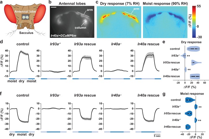
Figure 5. IR-dependent physiological responses to dry air.
(a) Schematic of the Drosophila head (viewed from above) illustrating the projection of IR40a/IR93a/IR25a-expressing neurons (green) (labeled using Ir40a-Gal4 [Silbering et al., 2011]) from the sacculus to the antennal lobes in the brain, visualized through a hole in the head cuticle. (b) Raw fluorescence image of Ir40a axons (in Ir40a-Gal4;UAS-GCaMP6m animals) innervating the arm and column in the antennal lobe. The dashed circle indicates the position of the ROI used for quantification in panels (d–g). (c) Color-coded images (reflecting GCaMP6m fluorescence intensity changes) of IR40a neuron responses to a switch from 90% to 7% RH ('Dry response') and to a switch from 7% to 90% RH ('Moist response'). (d,f) Moisture-responsive fluorescence changes in the arm (moist = 90% RH, dry = 7% RH). Traces represent average ± SEM. (e,g) Quantification of changes in ∆F/F (mean fluorescence change in the ROI shown in [b]) upon shift from moist to dry (e) or dry to moist (g). Dry responses were quantified as [∆F/F at 7% RH (average from 4.5 to 6.5 s after shift to 7% RH)] - [∆F/F at 90% RH (average from 3.5 to 1 s prior to shift to 7% RH)], and moist responses quantified by performing the converse calculation. Genotypes: control: n = 17 animals (pooled data from Ir40a-Gal4,Ir40a1/IR40a-Gal4,+;UAS-GCaMP6m/+, n = 9; IR40a-Gal4;UAS-GCaMP6m,Ir93aMI05555/+, n = 8). Ir93a mutant (Ir40a-Gal4;UAS-GCaMP6m,Ir93aMI05555/Ir93aMI05555), n = 10. Ir93a rescue (Ir40a-Gal4;UAS-GCaMP6m,Ir93aMI05555/UAS-mcherry:Ir93a,Ir93aMI05555), n = 8. Ir40a mutant (Ir40a-Gal4,Ir40a1;UAS-GCaMP6m/+), n = 8. Ir40a rescue (Ir40a-Gal4,Ir40a1;UAS-GCaMP6m/UAS-Ir40a), n = 6. **p<0.01, distinct from controls and rescues, Steel-Dwass test.

