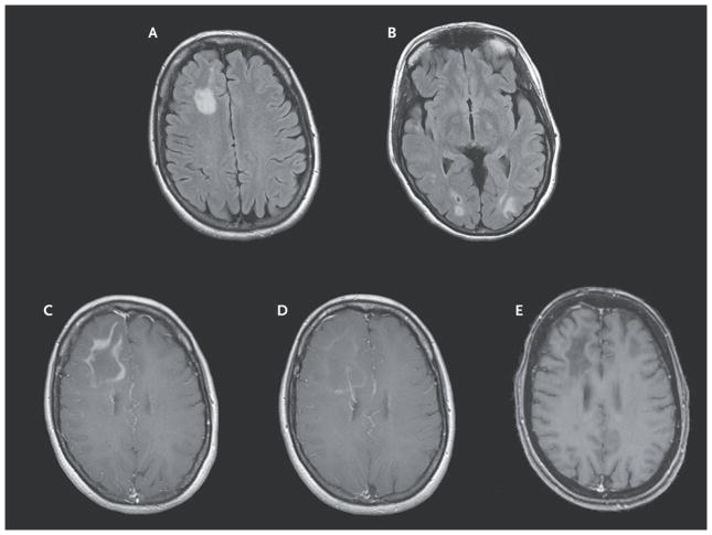
Figure 1. Sequential Magnetic Resonance Images of the Brain.
When the patient presented for treatment, axial fluid-attenuated inversion recovery images showed multilobar progressive multifocal leukoencephalopathy (Panels A and B). A T1-weighted image after gadolinium infusion showed frank immune reconstitution inflammatory syndrome (IRIS) after the patient discontinued maraviroc (Panel C).
After the patient began to receive maraviroc again, a T1-weighted image after gadolinium infusion showed regression of IRIS (Panel D). A T1-weighted image after gadolinium infusion obtained 2 months after gradual discontinuation of maraviroc showed stable lesions with no enhancement (Panel E).

