Figure 1.
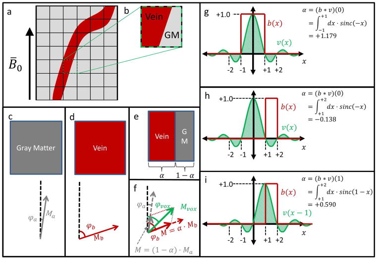
(a) A small vessel relative to the acquisition voxel size will suffer from partial-volume effects. (b) Such a voxel with partial-volume effects overlays two distinct tissue regions: vein and parenchyma (eg gray matter). (c) A voxel containing entirely gray matter has a complex signal with magnitude Ma and phase φa. (d) A voxel containing entirely venous blood has a complex signal with magnitude Mb and phase φb. (e) A voxel containing regions of each tissue consists of a fraction α of blood, and a fraction 1 − α of gray matter. (f) The partial-volume voxel signal will be the sum of the signals in (c) and (d) weighted by the fractions in (e). φa, φb, Ma, Mb, α, φvox, and Mvox are defined in Table 1. (g) Simplified 1D example where the vessel binary mask b(x) coincides with the main lobe of the voxel function v(x), and α > 1. (h) 1D example where b(x) coincides with a negative lobe of v(x), and α < 0. In (g) and (h), the different limits of integration represent the different spatial extents of the respective blood vessels. (i) For the same system as in (h), an adjacent voxel has positive α with a larger magnitude.

