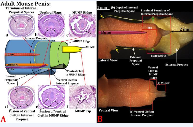
Figure 5.
(A) Diagram indicating the positions of internal and external structures within the mouse penis with histological sections (a-f) at the position specified. In section (a) the arrows indicate the proximal terminus on the epithelium defining the internal preputial space, whose position is indicated by the red arrow meeting the yellow line. In section (b) the arrows indicate the urethral flaps. In section (c) the arrows denote the distal aspect of the MUMP ridge with the MUMP located dorsal to the MUMP ridge. Section (d) the arrow denotes the fusion of the ventral cleft in the internal prepuce and thus stromal confluence across the midline. The red arrow indicates the position of this event on the diagram. The double-headed arrows indicate the internal prepuce. Section (e) denotes the fusion of the ventral cleft in the MUMP ridge and thus stromal confluence across the midline. The red arrow indicates the position of this event on the diagram. The doubleheaded arrows indicate the MUMP ridge. Section (f) depicts the bifid tips of the MUMP. In all cases the red arrows indicate where sections (a-f) are located on the diagram. (B) Photographs of penises of 60-day untreated mice in lateral and ventral views showing how the morphometric measures were made. (a) MUMP length = distance from MUMP tip to where the MUMP joins the MUMP ridge. (b) Depth of the internal preputial space was measured from the most distal section containing the internal prepuce proximally to the last section containing epithelium of the internal preputial space. (c) Internal preputial cleft length was measured from the most distal section containing the internal prepuce proximally to the proximal end of the cleft in the internal prepuce. The last measure (not illustrated) is distal penile tip to urethral flaps, that is, distal most section containing the tip of the MUMP to the first section containing urethral flaps. In the lateral view “Proximal Terminus of Internal Preputial Space” and “Bone Depth” indicate the surrogates of penile length used to normalize data. The upper image in (B) shows the external prepuce and the os penis (bone) superimposed. Yellow dotted lines indicated the full extent of the external prepuce, and the yellow arrows denote the reflection of the epithelium of the external prepuce onto the penile surface. Green arrows denote the point where the outer skin of the external prepuce becomes continuous with abdominal skin.

