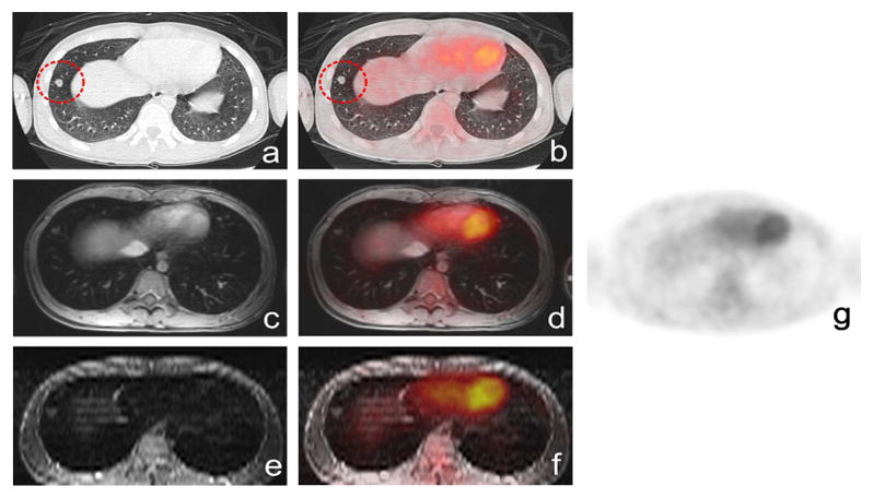
Fig. 3.

False positive, biopsy proven normal hypercellular hematopoietic marrow in a 15-year-old patient with lymphoma: (a) coronal ferumoxytol-enhanced FSPGR(b) coronal STIR scans show homogenous negative (dark) ferumoxytol-enhancement, confirming normal marrow, (c) hypermetabolic hematopoietic bone marrow on 18F-FDG PET and (d) on coronal 18F-FDG PET/FSPGR (ellipse), (e) hypermetabolic hematopoietic bone marrow on fused 18F-FDG-PET/STIR exams.
