. 2016 Sep 21;59(9):374–380. doi: 10.3345/kjp.2016.59.9.374

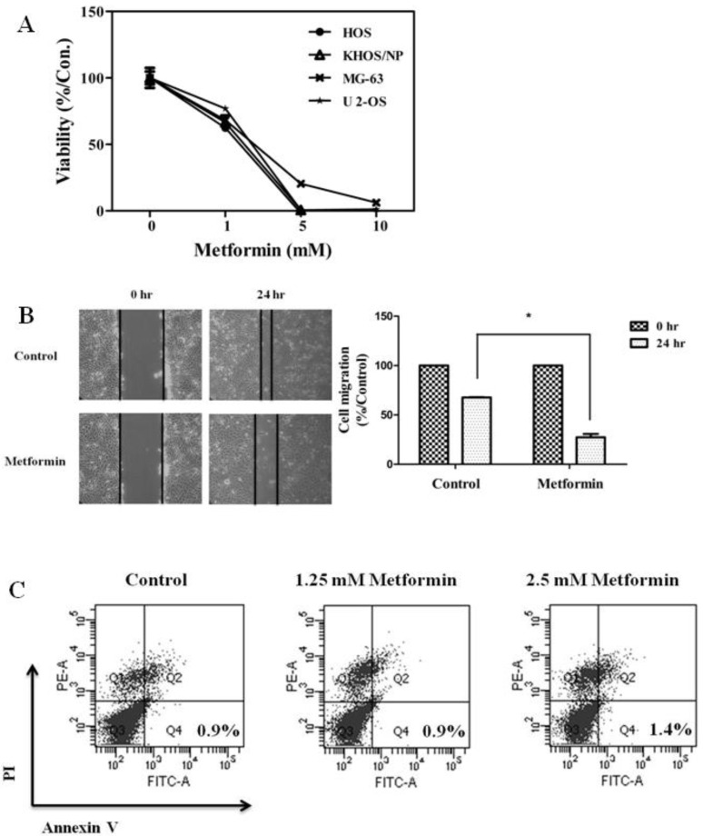
Fig. 1. (A) Metformin decreased the proliferation and migration of osteosarcoma cells. Four osteosarcoma cells were treated with metformin (0–10 mM) and cell viability assay (3-[4,5-dimethylthiazol-2-yl]-2,5-diphenyltetrazolium bromide) was performed 72 hours later. (B) KHOS/NP cells were plated in 6-well plates and grown to 80% confluence. After scratching, cells were incubated with or without metformin (1.5 mM). Microscopic appearance was captured 24 hours after wounding. The area of the gap region was quantified by ImageJ and data are shown in the bar graph (*P<0.05). (C) KHOS/NP cells were treated with metformin (1.25 or 2.5 mM) for 24 hours. Apoptotic cells were detected by Annexin V-propidium iodide (PI) dual staining.

Fig. 1