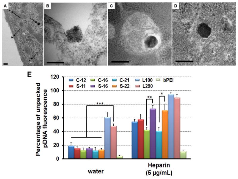
Figure 4.

Transmission electron microscopy of polyplexes form by S-22 interacting with HeLa cells, including overview (A), endocytosis (B), entrapped in endosome (C) and locating in cytoplasm (D). HeLa cells were treated with polyplexes for 1 h prior to fixation and preparation for TEM. Scale bar indicates 200 nm. (E) Unpacking study of polyplexes (N/P = 5) in the absence and presence of heparin sulfate (5 μg/mL). Data are shown as mean ± SD (n = 3; student's t-test, *p < 0.05, **p < 0.01, ***p < 0.001).
