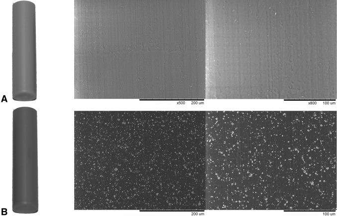
Fig. 1A–B.
Macroscopically, PEEK-HA (B) appears slightly gray compared with PEEK (A). Backscattering scanning electron microscopy at ×100 and ×500 demonstrates comparable surface topography and the presence of the HA incorporated into PEEK-HA that is present on the surface of the material and appears as white particulate under electron microscopy.

