
FIGURE 2.
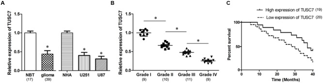
(A) Compared with normal brain tissues (NBT) and normal human astrocytes (NHA) cells, TUSC7 was poorly expressed in human glioma tissues and cell lines. (B) Glioma tissues with worse histological grade exhibited decreased TUSC7 expression. (C) Kaplan–Meier analysis indicated that lower TUSC7 expression was associated with a significantly shorter survival time than higher TUSC7 expression. Data are presented as mean ± SD (n = 5, each group). ∗P < 0.05 vs. NBT or NHA group.