Fig. 2.
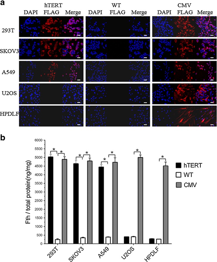
Analysis of FLAG-tagged Fth expression in cells transfected with or without lentiviral vector. a Immunofluorescence staining of cells with anti-FLAG antibody. After transfection with Lenti-hTERT-Fth1-3FLAG-Puro, FLAG-tagged protein was identified in immunofluorescence images of A549, SKOV3 and 293T cells but not U2OS or HPDLF cells. Red fluorescence represents FLAG-tagged protein; blue fluorescence represents cell nuclei. Scale bar 50 μm. b ELISA for Fth protein in all cell types. In A549, SKOV3 and 293T cells, but not U2OS or HPDLF cells, transfected with Lenti-hTERT-Fth1-3FLAG-Puro, Fth protein level was significantly increased compared to the untransfected cells. Error bars indicate standard deviations of triplicate samples. *p value was statistically significant (p < 0.05). hTERT Lenti-hTERT-Fth1-3FLAG-Puro-transfected cells, CMV Lenti-CMV-Fth1-3FLAG-Puro-transfected cells, WT wild-type cells

