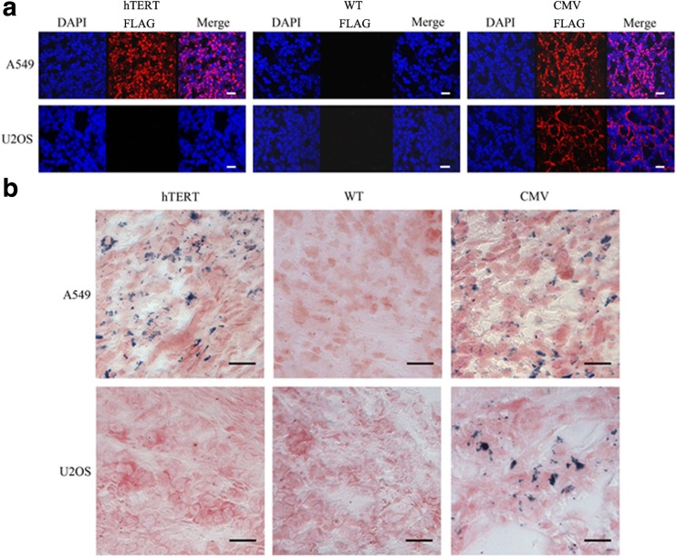
Fig. 6.
Histological analyses of tumour sections. a Immunofluorescence staining of FLAG protein in tumours with anti-FLAG antibody. Red fluorescence represents FLAG-tagged proteins; blue fluorescence represents cell nuclei. Scale bar 50 μm. b Prussian blue staining of iron deposits in tumour tissues. Scale bar 10 μm. hTERT Lenti-hTERT-Fth1-3FLAG-Puro-injected tumours, CMV Lenti-CMV-Fth1-3FLAG-Puro-injected tumours, WT wild-type tumours

