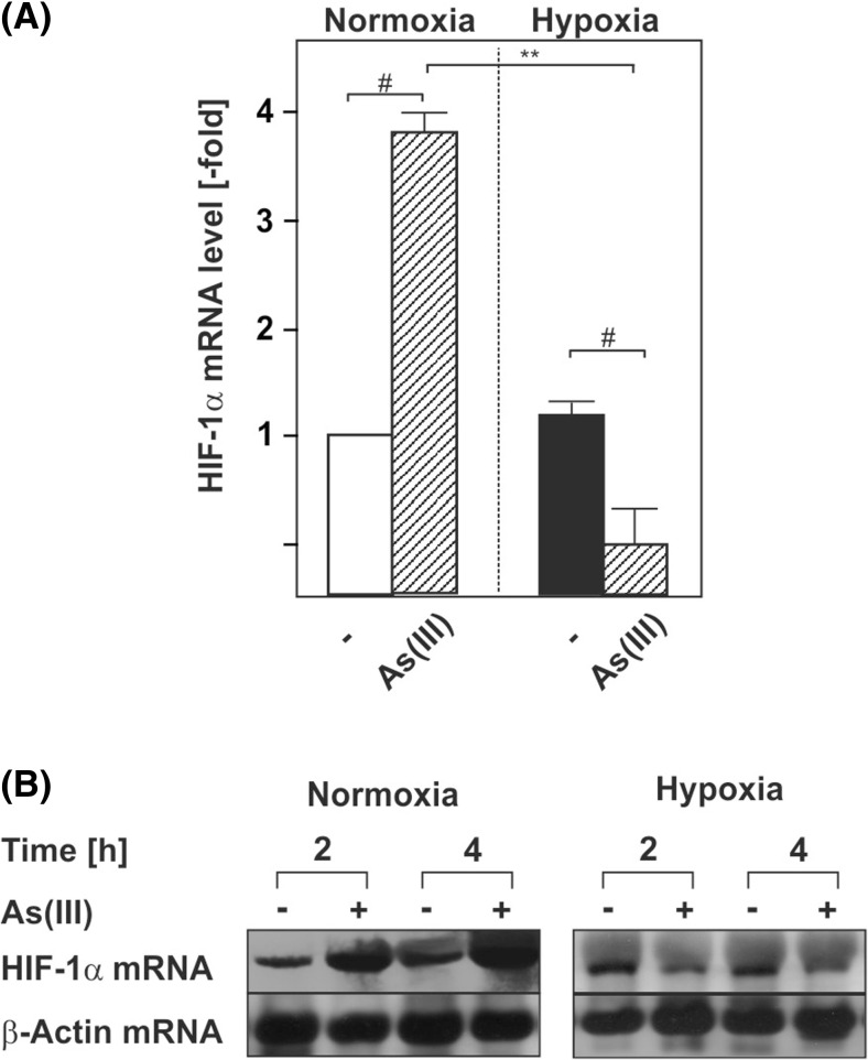
Fig 2.
As(III) induces HIF-1α at the transcriptional level. Cells were cultured under normoxia for 24 h. After 24 h, the medium was changed and cells were further cultured under normoxia and hypoxia in the absence or presence of 50 μM As(III). a, b The HIF-1α mRNA expression levels under normoxia (16 % O2) were set to 1. Values represent means ± SEM of three independent experiments. Single number sign, significant difference As(III) vs. respective control; double asterisks, significant difference As(III) at 16 % O2 vs. As(III) at 5 % O2; p ≤ 0.05. b Representative Northern blot. Twenty micrograms of total RNA from cultured HepG2 cells were subjected to Northern blot analysis and hybridized with DIG-labeled HIF-1α and β-actin probes. Autoradiographic signals were visualized by chemiluminescence and quantified by video densitometry

