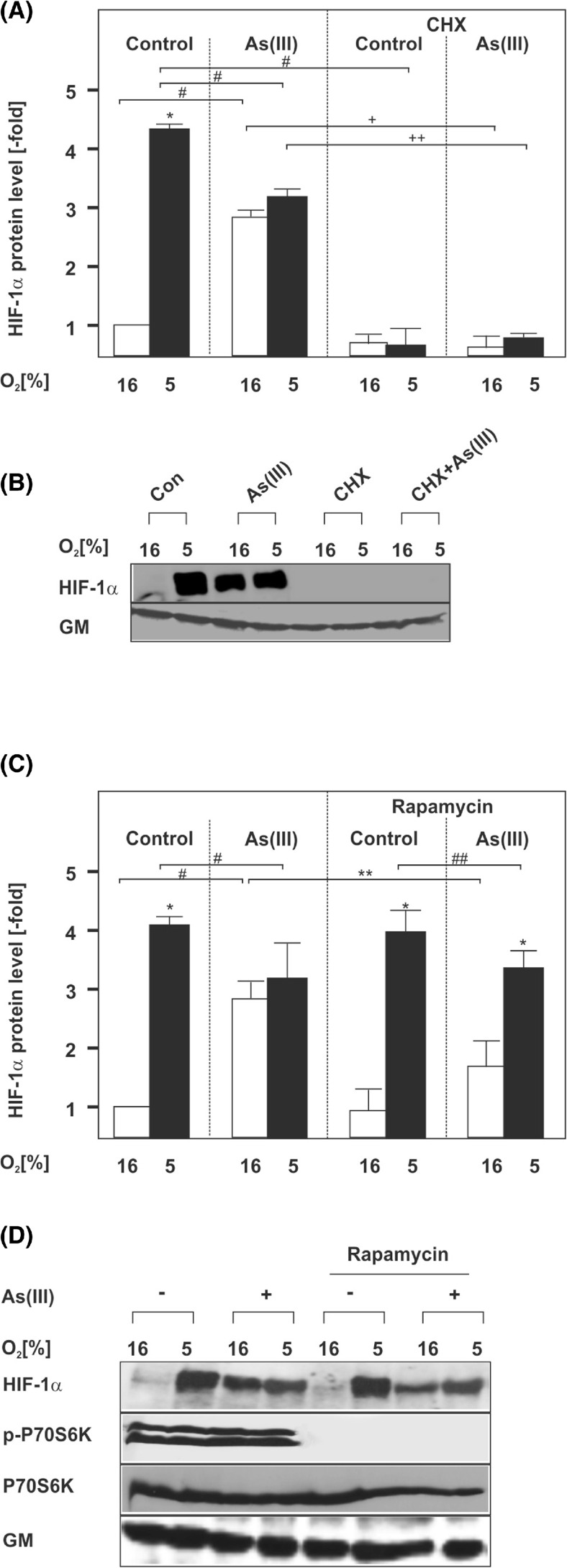
Fig. 3.
Induction of HIF-1α by As(III) is dependent on mTORC1-driven protein synthesis. Cells were cultured under normoxia for 24 h. At 24 h, the medium was changed and the cells were pre-treated for 30 min either with cycloheximide (CHX, 10 μg/ml) (a, b) or rapamycin (10 μM) (c, d) and then treated with 50 μM As(III) under normoxia (16 % O2) or hypoxia (5 % O2) for 4 h. a, c Statistical analyses of HIF-1α levels. The HIF-1α levels in the controls were set to 1. Values represent mean ± SEM of three independent experiments. Single asterisk, significant difference 16 % O2 vs. 5 % O2; single number sign, significant difference As(III) vs. control; double asterisks, significant difference 16 % O2 + As(III) vs. rapamycin at 16 % O2 + As(III); double number sign, significant difference rapamycin at 5 % O2 vs. rapamycin at 5 % O2 + As(III); single plus sign, significant difference As(III) 16 % O2 vs. As(III)/CHX 16 % O2; double plus sign, significant difference As(III) 5 % O2 vs. As(III)/CHX 5 % O2; p ≤ 0.05. b, d Representative Western blot. One hundred micrograms of isolated total protein was subjected to Western blot analysis with an antibody against HIF-1α, Golgi membrane (GM), phospho-p70S6K1, or total p70S6K1. Signals were visualized by chemiluminescence and quantified by video densitometry

