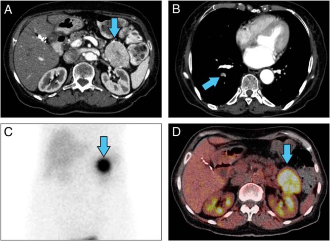
Figure 1.
Radiographic imaging of a patient with VIPoma. A, Contrast-enhanced CT scan of the abdomen showed pancreatic tail mass (blue arrow). B, A pulmonary CT angiography showed right segmental pulmonary artery-filling defects. C, Octreoscan. D, F18-FDG PET/CT scan showed an intense uptake in the pancreatic tail mass.

