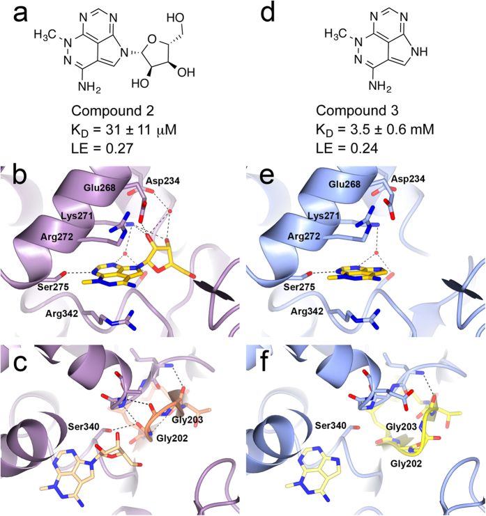
Figure 1. Binding mode of triciribine.
(a) Chemical structure of the AKT inhibitor triciribine 2. (b) Structure of 2 (yellow) bound to HCS70-NBD/BAG1 (purple). (c) Structure of HSC70-NBD/BAG1 (purple) bound to triciribine (beige) showing the regular conformation of the phosphate-binding loop 2 (orange). (d) Chemical structure of triciribine fragment 3. (e) Structure of 3 (yellow) bound to HCS70-NBD/BAG1 (blue). (f) Structure of HSC70-NBD/BAG1 (blue) bound to 3 (light yellow) showing the flexibility in the phosphate-binding loop 2 (yellow) which disrupts the phosphate-binding pocket. LE = ligand efficiency50.

