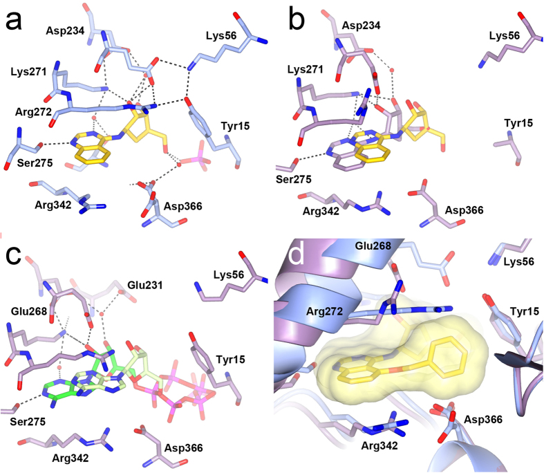
Figure 5. NBD flexibility and aminoquinazoline binding.
(a) Structure of 23 (yellow) bound to the HSP72-NBD (light blue). (b) Superposition of open HSC70-NBD/BAG1 and closed HSP72-NBD 23-bound structures showing the shift in compound position. Key HSC70-NBD protein residues and the HSC70-bound compound 23 are displayed in purple and superimposed HSP72-NBD bound compound 23 in yellow. (c) Superposition of the ATP-bound HSC70-BAG1 structure (PDB ID 3FZF) in the open conformation with the ATP-bound HSP72-NBD in the closed conformation showing the shift in the nucleotide. The bound ATP molecule is displayed in green and light green in the respective structures. HSP72-NBD residues are omitted for clarity in panel b and c. (d) Superposition of the HSP72-NBD structures bound to 23 and 28 showing the effects of the 5-O-benzyl substituent. The 28-bound HSP72-NBD is shown in purple and the 23-bound HSP72-NBD in light blue. Compound 28 is shown in yellow with a transparent molecular surface superimposed. Compound 23 is omitted for clarity.

