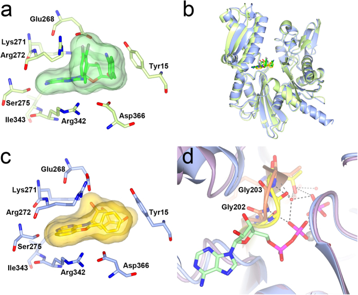
Figure 6. Comparison with VER-155008 (1) and conformational flexibility in the phosphate binding loop 2.
(a) Close-up of VER-155008 (1, green) bound to the HSP72 NBD (light-green PDB ID: 4IO8). The compound surface is shown as a semi-transparent green surface. (b) Superposition of the 1-bound (light green) and 28-bound (blue) HSC72-NBD structures showing that the latter is in a more, but not completely, closed conformation. The ligands are displayed in green and yellow. (c) Close-up of the 28 (yellow) bound to the HSP72-NBD structure showing its 5-O-benzyl substituent occupies a similar area as the substituents of 1, but interacts in a different way. The compound surface is shown as a semi-transparent yellow surface. (d) Superposition of the nucleotide-free HSC70-NBD/BAG1 structure (PDB ID 1HX1, purple) and the ADP/Pi bound HSP72 structure (PDB ID 1S3X, light blue) showing the effect of the phosphate-binding loop 2 conformations on its interactions with ADP (green). The respective conformations of the phosphate-loop 2 are shown in yellow and orange.

