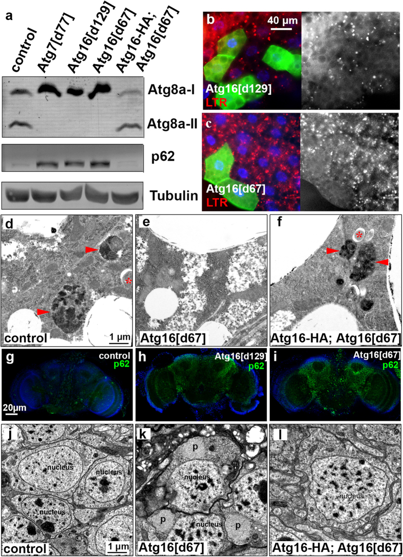
Figure 2. Atg16 is required for autophagy in larval fat and adult brain cells.
(a) Lipidated, autophagosome-associated Atg8a (Atg8a-II) is missing and the selective autophagy cargo p62 accumulates in Atg16d129 and Atg16d67 mutant larval lysates, similar to Atg7d77 mutants. These phenotypes are rescued by Atg16-HA expression in Atg16d67 mutants. (b,c) Starvation-induced punctate LTR staining is blocked in homozygous Atg16d129 (b) and Atg16d67 (c) mutant fat body cells that are marked by GFP expression, compared to neighboring GFP-negative control cells. (d–f) Ultrastructural images of fat cells in starved L3 larvae. Autophagosomes (asterisks) and autolysosomes (arrowheads) are seen in control (d) and genetically rescued (f) animals, unlike in Atg16d67 mutant fat body cells (e). (g–i) Endogenous p62-positive protein aggregates (green dots) accumulate throughout the brain of Atg16d129 (h) and Atg16d67 (i) animals compared to control brain (g). (j–l) Ultrastructural analysis of 20-day-old brains reveals the presence of large protein aggregates (p) in Atg16d67 mutant neurons (k), which are absent from control (j) or genetically rescued (l) animals.

